
How to design biologically meaningful droplet assays for single cells, enzyme engineering, and microbial discovery—and what to measure before scaling a workflow.
Microfluidic droplets make it possible to partition cells, enzymes, and chemical reactions into thousands of isolated microscale compartments. That physical separation can reveal responses that are obscured in bulk samples, reduce reagent consumption, and connect an individual cell or genetic variant to a measurable phenotype. Yet producing uniform droplets is only one part of a useful screening platform. Researchers must also control sample loading, biological compatibility, reaction timing, signal detection, and recovery of selected compartments.
The most productive question is therefore not simply “How many droplets can the chip make?” but “How reliably does each droplet generate an interpretable biological result?” From prototype design to assay validation, a practical strategy combines custom microfluidic encapsulation services with appropriate flow control, application-specific sample handling, and robust analytical readouts.
Why Droplet Encapsulation Changes the Screening Experiment
In conventional bulk assays, secreted enzymes, metabolites, and signaling molecules may diffuse throughout the sample. Droplet microfluidics changes that relationship by creating confined aqueous reaction volumes within an immiscible carrier phase. A cell and its molecular products can remain associated long enough for a detectable response, provided the reporter, emulsion chemistry, and incubation conditions are compatible.
A modular droplet-screening workflow may combine encapsulation, incubation, reagent addition, fluorescence detection, and sorting. Different studies implement these functions with different device designs, so the value of the method lies in connecting the modules around a defined biological endpoint—not assuming one chip architecture will work for every specimen.
The approach is especially relevant for heterogeneous microbial populations, enzyme-variant libraries, and cell-based compound screens. However, throughput does not automatically equal information quality: empty droplets, double occupancy, unstable emulsions, assay cross-talk, and selection bias can all erode the value of a large droplet count.
Designing an End-to-End Microfluidic Screening Workflow
1. Define the biological question before choosing the chip
Start with the unit of analysis. Is the goal to identify one enzyme-producing microorganism, rank a pool of engineered protein variants, characterize individual mammalian cell responses, or examine a microbial consortium? A single-cell study may require a low probability of more than one cell per droplet; a co-culture experiment may intentionally use two or more organisms. In contrast, bead- or hydrogel-based workflows may prioritize scaffold architecture rather than a free water-in-oil compartment.
When cells enter droplets randomly at an average loading rate λ, occupancy is often approximated by a Poisson distribution: P(k) = e^(−λ) λ^k / k!. Reducing λ lowers the frequency of droplets with multiple cells but increases the number of empty droplets. Consequently, “high single-cell purity” and “high fraction of occupied droplets” are distinct objectives. Ordered loading, upstream cell focusing, or downstream enrichment may be valuable when sample scarcity makes excessive empty droplets unacceptable. For projects requiring compartment-level loading optimization, microfluidic cell encapsulation solutions provide an application-relevant entry point.
2. Match droplet geometry to the sample and readout
Flow-focusing junctions and T-junction geometries commonly generate droplets by shearing a dispersed aqueous phase with an immiscible continuous phase. Droplet volume, size distribution, and formation frequency depend on channel geometry, flow-rate ratio, interfacial tension, fluid viscosity, and wetting properties. A strong assay design treats these as interacting variables rather than optimizing one flow rate in isolation.
Microscopy-based sizing, stable flow-rate monitoring, and observation of breakup modes are useful early checks. Customized flow-type analysis chips and droplet generator and flow-chemistry solutions can support the development of architectures suited to the intended droplet size, sample composition, and downstream module.
3. Protect the biological payload
Carrier oil and surfactant selection is as important as geometry. A stable emulsion must preserve droplet integrity while maintaining sufficient oxygen availability, viability, enzyme activity, or analyte retention for the assay. The optimal chemistry depends on incubation duration and the biological material; it should be tested rather than assumed biocompatible. Small hydrophobic compounds and some fluorescent reporters may partition between aqueous droplets and oil or migrate through surfactant interfaces, creating apparent biological cross-talk.
Where prolonged culture or controlled mechanical support is essential, hydrogel particle synthesis or other microgel approaches may provide an alternative format. Researchers interested in microcapsules and formulations can also explore microfluidics-based particle synthesis services, although droplet synthesis and a validated live-cell assay are not interchangeable deliverables.
4. Coordinate incubation, reagent addition, and detection
Some functional assays can be formed in one step by co-encapsulating cells, substrates, and reporters. Others require staged addition after growth or expression. Droplet merging and picoinjection can introduce assay reagents at a chosen time, but both demand consistent dosing and compatibility with the emulsion. A short reaction may favor on-chip residence-time control, whereas a culture-dependent endpoint may require stable off-chip incubation and reinjection.
Optical readouts such as fluorescence are widespread in droplet screening because intensity can be interrogated rapidly. Nevertheless, fluorescence must be normalized or controlled for droplet size, substrate availability, reporter leakage, autofluorescence, and changes in cell number. Integration with microfluidic solution mixing chip development may help standardize reagent introduction upstream or in auxiliary channels; a broader microfluidic biosensor development workflow may be more appropriate when the endpoint is not compatible with fluorescence.
5. Sort, recover, and revalidate
A successful screen should define how selected droplets become usable biological material or analyzable evidence. Fluorescence-activated droplet sorting can enrich compartments that exceed a chosen signal threshold. Depending on the experiment, recovery may involve breaking emulsions, regrowing cells, sequencing isolates, or repeating functional measurements under independent conditions. The selected phenotype should be confirmed after recovery, because the screening environment may not fully reproduce the conditions of a downstream application.
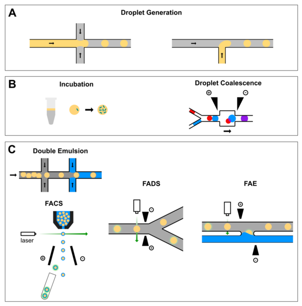
Fig. 1 A typical workflow of droplet-based high-throughput screening system for protein engineering.1,3
What Can Be Screened? Three Practical Use Cases
Enzyme activity and protein engineering
For an enzyme engineering campaign, a droplet can link the activity of an enzyme variant to its encoding cell, gene, or reaction compartment. A fluorogenic substrate generates a measurable signal, and high-performing droplets may then be enriched for secondary characterization. The screening system must preserve genotype–phenotype linkage: a variant cannot be interpreted confidently if enzyme or product readily escapes into neighboring droplets.
Assay design should consider substrate conversion kinetics, signal linearity, incubation temperature, reagent toxicity, and the likelihood that a bright signal reflects faster catalysis rather than greater cell growth or expression. A follow-up assay under relevant process conditions is essential before assigning superiority to an enzyme hit. Related service areas include enzyme encapsulation and customized compartmentalization for biochemical screening.
Single-cell responses and heterogeneous populations
Single-cell droplet assays can measure variation in reporter activity, secreted factors, viability, or other droplet-compatible signals. They are particularly useful when population averages conceal rare but biologically important subgroups. The experimental unit must be carefully defined: one droplet is not necessarily one viable cell, and one fluorescence reading is not necessarily an independent measure of biological function.
For cell-based drug studies, exposure duration, compound partitioning, cell-cycle state, optical interference, and post-encapsulation survival should be measured alongside response endpoints. A careful feasibility assessment can distinguish experiments well served by single-cell microencapsulation from applications better addressed by culture chambers, hydrogel carriers, or non-droplet microfluidic platforms.
Cultivation and recovery of functional gut bacteria
Droplet isolation can also make microbial cultivation more experimentally tractable. In a 2022 study, Yin and colleagues used a droplet-based workflow to isolate gut bacteria that could utilize metabolites associated with an engineered butyrate-producing bacterium. The researchers generated single-bacterium droplets, spread them on agar, and cultured the material under anaerobic conditions. Their approach illustrates how microfluidic compartmentalization can be connected to familiar downstream cultivation and isolate-recovery steps, rather than requiring every operation to be performed on chip.
For a microbiome project, the crucial decisions include sample preprocessing, clumping and cell concentration, medium composition, aerobic or anaerobic handling, droplet integrity during transfer, and the criteria for “functional” enrichment. Culturing a recovered organism does not in itself establish clinical benefit or therapeutic suitability. The workflow should include independent identification and functional confirmation of isolates. Creative Biolabs lists related capabilities in bacteria encapsulation and microbiome encapsulation.
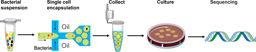
Fig. 2 Schematic of the workflow using the single-cell droplet culture approach to search for functional bacteria from gut microbiota.2,3
Where Microfluidic Modules Add Value Beyond Droplet Generation
Not every microfluidic application requires discrete droplets. A laminar flow control platform can provide controlled interfaces for sample handling; gradient-generating microfluidic chips can establish reproducible concentrations for chemotaxis, dose-response, or environmental exposure studies; and integrated microfluidic biosensors can provide electrical or optical outputs. These approaches may complement a droplet screen, or replace it when long-term imaging, surface attachment, or continuous-flow measurement is the priority.
A modular project plan therefore begins with the analyte, biological question, throughput target, and downstream readout. It then selects the device architecture, materials, and controls that fit the question. This helps avoid an attractive prototype that generates droplets successfully but cannot produce robust, actionable data.
How Creative Biolabs Can Support a Custom Microfluidic Project
Creative Biolabs provides microfluidic development and encapsulation-related services spanning device design and workflow customization. Depending on project requirements, relevant capabilities include custom encapsulation solutions, droplet generation and flow-chemistry development, flow analysis chip development, solution mixing chip development, enzyme encapsulation, and microbiome encapsulation. The appropriate combination depends on the biological payload, target microenvironment, assay duration, and required analytical endpoint.
Researchers considering a custom platform can start by sharing the sample type, approximate target droplet size or payload, proposed readout, necessary incubation period, expected throughput, and recovery requirements. An early technical discussion can help identify which parameters must be validated before investing in a fully integrated device. Explore Creative Biolabs’ microfluidics capabilities or discuss a tailored microfluidic encapsulation workflow to assess a project-specific strategy.
References
- Weng, Lindong, and James E. Spoonamore. “Droplet Microfluidics-Enabled High-Throughput Screening for Protein Engineering.” Micromachines, vol. 10, no. 11, 2019, article 734. https://doi.org/10.3390/mi10110734
- Yin, Jianan, et al. “A Droplet-Based Microfluidic Approach to Isolating Functional Bacteria from Gut Microbiota.” Frontiers in Cellular and Infection Microbiology, vol. 12, 2022, article 920986. https://doi.org/10.3389/fcimb.2022.920986
- Distributed under Open Access license CC BY 4.0, without modification.