Cancer remains a formidable challenge due to its difficulty in detection. It has become the second leading cause of death worldwide. The main reasons for the poor efficacy of cancer treatment include drug resistance and treatment variability caused by cancer cell heterogeneity. In addition, drug screening using cancer cells based on two-dimensional (2D) culture fails to consider the impact of the in vivo tumor microenvironment, leading to inaccurate evaluation of drug efficacy. Therefore, single-cell analysis of tumor cells and the development of in vitro tumor models are urgent issues in cancer research. After decades of development, droplet microfluidics technology has been widely used in single-cell analysis and three-dimensional (3D) cell culture, providing a new and powerful platform for the heterogeneity analysis of tumor cells and in vitro tumor spheroid culture.
Recently, a review entitled “Droplet Microfluidics for current cancer research: from single-cell analysis to 3D cell culture” was published in the journal ACS Biomaterials Science & Engineering, outlining the latest progress and application prospects of droplet microfluidics in current cancer research.
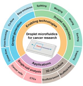
This review first outlines the enabling technologies of droplet microfluidics, including the generation of single/double emulsion droplets and the active/passive splitting, fusion, mixing, sorting, and capture of droplets. Then, the use of droplet microfluidics for the heterogeneous analysis of single-cell genomics, transcriptomics, proteomics, and metabolomics is systematically discussed, and the application of the combination of droplet microfluidics and microfluidic sorting technology in single-cell analysis of rare circulating tumor cells is emphasized. In addition, the paper discusses the scaffold-free 3D tumor spheroid culture based on droplet microfluidics technology, as well as the strategy of using different hydrogels in droplet microfluidics technology to achieve scaffold-based 3D tumor spheroid culture. Finally, the paper discusses the challenges and development prospects of the application of droplet microfluidics in the field of cancer research.
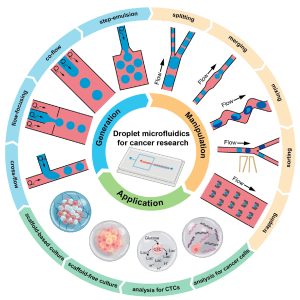 Figure 1. Application of Droplet Microfluidics Technology in Cancer Research
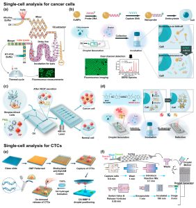 Figure 2. Droplet microfluidics for cancer cell research: (a) single-cell CD-45 transcriptome analysis; (b) single-cell miRNA-21 analysis; (c) single-cell secreted vascular endothelial growth factor analysis; (d) single-cell secreted alkaline phosphatase analysis. Droplet microfluidics for rare circulating tumor cells: (e) combining antibody-based cell capture and acoustic droplet technology to achieve genomic mutation analysis of rare circulating tumor cells; (f) combining size-based cell capture and step emulsification to achieve matrix metalloproteinase metabolic analysis of rare circulating tumor cells.
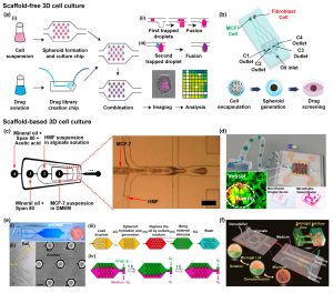 Figure 3. Droplet microfluidics for scaffold-free 3D tumor spheroid culture: (a) Ewing sarcoma spheroid culture and drug resistance testing; (b) Culture and drug resistance testing of tumor spheroids containing different ratios of MCF-7 breast cancer cells and NIH-3T3 fibroblasts. Droplet microfluidics for scaffold-based 3D tumor spheroid culture: (c) Alginate microcapsules encapsulating MCF-7 breast cancer cells and HMF breast fibroblasts to achieve 3D tumor spheroid culture; (d) GelMA droplets for vascular-supported multicellular tumor spheroid culture; (e) Agarose droplets for long-term culture, drug perfusion, and on-demand extraction of tumor spheroids; (f) Integrated chips for long-term culture of large quantities of tumor spheroids.
In summary, droplet microfluidics has become a powerful platform for single-cell analysis and 3D cell culture, offering low cost, high sensitivity and high throughput. It provides a valuable tool for analyzing the heterogeneity of tumor cells and cultivating in vitro tumor models. Nevertheless, there are still challenges and development prospects for further application of droplet microfluidics in cancer research.
The introduction of machine learning and artificial intelligence can solve the current reliance on researchers’ experience for designing droplet microfluidic chips. Additionally, combining label-free detection methods, such as image recognition enabled by artificial intelligence, can broaden the scope of droplet-based single-cell analysis. On the other hand, advancements in rapid microfabrication technologies, such as 3D printing, can overcome the issues of long manufacturing cycles and high costs associated with traditional soft lithography. Developing hydrogels with excellent biocompatibility and controllable physical and chemical properties can better simulated the in vivo tumor microenvironment, facilitating the culture of in vitro tumor spheroids.
Reference:
Jiang L, Guo K, Chen Y, Xiang N. Droplet Microfluidics for Current Cancer Research: From Single-Cell Analysis to 3D Cell Culture. ACS Biomater Sci Eng. 2024;10(3):1335-1354. doi:10.1021/acsbiomaterials.3c01866
Related Services:
3D Tissue and Organ-On-A-Chip Model Development Services
Single Cell Sequencing Microfluidic Chip Development Service