A new microfluidic technology has recently emerged, revolutionizing the extraction of minuscule extracellular vesicles (sEVs) from human blood. Scientists at ETH Zurich, Switzerland, spearheaded this research, unveiling a straightforward viscoelastic-based microfluidic platform. This innovation allows the efficient and label-free isolation of sEVs from human blood, boasting superior separation effects and significantly augmenting sEV yields in comparison to traditional ultracentrifugation methods. In terms of practical clinical applications, the research team successfully isolated sEVs from both cancer patients and healthy donors, unveiling higher concentrations of sEVs in the blood of cancer patients. Their groundbreaking findings were published in the esteemed academic journal Science Advances on October 6 titled “Direct Isolation of Small Extracellular Vesicles From Human Blood Using Viscoelastic Microfluidics.”
Extracellular vesicles (EVs) are ubiquitous in bodily fluids, including blood, saliva, and urine. These lipid-based vesicles, produced by cells, encapsulate biologically active payloads like nucleic acids, proteins, and metabolites. Based on their size, EVs are classified into three subtypes: small (sEVs; <200 nm), medium (mEVs; 200 to 800 nm), and large (lEVs; >800 nm). Among these, sEVs are particularly significant as they serve as invaluable non-invasive diagnostic and prognostic biomarkers. However, traditional methods of sEV isolation, such as ultracentrifugation, gradient centrifugation, or microfiltration, suffer from several limitations, including prolonged long processing times, low yields (ranging from 5-40%), compromised integrity of isolated sEVs, and high equipment costs.
In the past decade, microfluidic technologies have gained prominence as fundamental tools for processing and isolating sEVs, owing to their precision in manipulating micron- and nanoscale objects. These microfluidic strategies can be broadly categorized as passive or active, depending on their mode of isolation. Passive methods leverage size-dependent hydrodynamic forces or intricate channel structures such as nanopore membranes and nanopillar arrays, eliminating the need for external forces. On the other hand, active methods necessitate external force fields, typically commonly acoustic, electric, or magnetic, to manipulate sEVs.
Active microfluidic techniques have successfully isolated sEVs from blood, demonstrating high separation efficiency. Through chip-based electrophoresis technology, scientists have captured sEVs smaller than 500 nm from whole blood and subsequently conducted on-chip immunofluorescence assays to detect these captured sEVs. However, this method faces challenges over extended periods due to decreasing separation efficiency caused by the accumulation of sEVs on the electrode array. Another approach involves the use of magnetic Fe3O4 nanoparticles functionalized with antibodies to capture sEVs from whole blood, particularly in the context of pancreatic cancer. Although effective, this method involves complex steps of antibody modification and conjugation, making it cumbersome for clinical settings.
Passive microfluidic systems, when combined with physical filtration, have also proven successful in isolating sEVs from whole blood. For instance, a multifaceted membrane-integrated microfluidic device can isolates sEVs (<200 nm) from small blood samples, utilizing a polycarbonate membrane for agitation-enhanced filtration. While this method is practical, its applicability is hindered by the intricate device structure and the requirement for pneumatically driven “micro-stirrs” to prevent device clogging.
In clinical settings, the pressing demand for cost-effective, user-friendly exosome (or EV) isolation methods necessitates techniques that require no specialized skills, minimal sample processing, and yet ensure high isolation purity, recovery, consistency and reproducibility. Passive microfluidic methods for isolating sEVs from whole blood offer distinct advantages in terms of affordability, ease of operation, and reproducibility. However, they are not without their challenges. Filtration-based platforms, for instance, are prone to membrane clogging. Conversely, passive technologies relying on inertial and viscoelastic microfluidics exhibit superior recovery and purity rates. In the clinical context, there is an urgent need for microfluidic tools that are efficient, straightforward, and affordable, capable of swiftly and consistently isolating sEVs from whole blood.
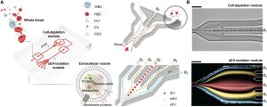
The research team harnessed microfluidic technology to develop a viscoelastic-based microfluidic device designed to efficiently extract sEVs from human blood. This innovative system comprises two modules: a cell removal module and an sEV isolation module. In the first module, micron-sized blood components such as white blood cells (WBCs), red blood cells (RBCs), and platelets (PLTs) are effectively eliminated from the blood sample, leaving behind a “cell-free” blood solution. This purified blood is then seamlessly introduced into the second module, where sEVs are meticulously separated and isolated from other EV subgroups.
The research findings underscore the device’s exceptional separation capabilities, achieving a remarkable purity rate of 97% and a recovery rate of 87% for fluorescently labeled sEVs isolated from whole blood. When compared to conventional ultracentrifugation (UC) methods, this microfluidic approach significantly enhances sEV yields. Notably, proteomic analysis revealed a striking similarity in the protein composition of sEVs extracted using both methods.
To validate the technology’s applicability, the research team extracted sEVs from blood samples obtained from 20 cancer patients and 20 healthy donors. The concentration and size of these sEVs were compared with data obtained through the gold standard ultracentrifugation method. The results unequivocally demonstrated substantially elevated sEV concentrations in the blood of cancer patients compared to healthy donors.
This groundbreaking research heralds a new era in sEV extraction and utilization. The technology, characterized by its simplicity, efficiency, and label-free nature, is poised to revolutionize clinical diagnosis and treatment methodologies. Looking ahead, the research team aims to refine this technology further and explore its potential applications in various diseases.
Published in the journal Science Advances, these research findings breathe new life into the realm of small extracellular vesicles, offering fresh optimism for both research and practical clinical applications. The successful development of this technology promises to deepen the understanding of sEVs and provide clinicians with more accurate and convenient diagnostic and treatment options.
Reference:
Meng Y, Zhang Y, Bühler M, et al. Direct isolation of small extracellular vesicles from human blood using viscoelastic microfluidics. Sci Adv. 2023;9(40):eadi5296. doi:10.1126/sciadv.adi5296
Related Services: