The intestine refers to the alimentary canal from the pylorus of the stomach to the anus, which is the longest section of the alimentary canal and is also an important digestive organ of the human body. The main functions of the human gut are the regulation of digestion, absorption, and secretion, and the establishment of a protective epithelial barrier between the human body and the digestive environment. In addition, the intestine is also an important immune organ, and the interaction between intestinal microorganisms, intestinal lymphoid tissue, and the host immune system constitutes the homeostasis of the intestinal environment.
For experimental research on the intestinal tract, traditional in vitro models rely on the static culture of intestinal epithelial cells, which lack the natural microenvironment, intraluminal fluid flow, and periodic peristalsis of the normal intestinal tract, and cannot support the co-culture between the commensal microbiome and human intestinal cells. While in vivo models such as germ-free mouse models have species differences from humans. In 2012, the Wyss Institute of Harvard University invented a human gut-on-a-chip for the first time (shown below). The gut chip is made of polydimethylsiloxane (PDMS) and consists of 2 parallel microchannels separated by a porous flexible membrane coated with extracellular matrix (ECM), lined with human intestinal epithelium (Caco- 2) cells. The complex structure and physiology of the living intestine can be imitated, and it allows the application of cyclic mechanical force to simulate the motor function of the intestine in vivo, which is of great significance for drug testing and the development of new intestinal disease models.
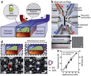 Schematic diagram of the human gut organ-on-a-chip
Intestinal Organ Chip Construction Technology
The construction of the intestinal microfluidic chip mainly includes five steps: making a microstructured silicon wafer template, making a PDMS membrane with a microstructure, making a PDMS microchannel layer, sealing the chip, and adding intestinal-related cells. The specific construction method is as follows:
Fabrication of Microstructured Silicon Wafer Templates
Before preparing the V-groove, the silicon wafer was cleaned by a standard RCA method, and a layer of SiO2 was evaporated on the surface of the silicon wafer by plasma enhanced chemical vapor deposition (PECVD). Then, photolithography was performed using a pre-designed photolithography plate to obtain a patterned SiO2-covered single-crystal silicon substrate. Subsequently, the substrate is etched anisotropically by using a mixed solution of tetramethylammonium hydroxide and isopropanol in a certain proportion. Finally, the SiO2 layer on the surface was removed using an oxide etchant to obtain the required patterned silicon template.
Fabrication of PDMS Microstructured Membranes
The patterned silicon template was placed in a vacuum desiccator, and the surface of the silicon membrane plate was modified by adding perfluorooctyltrichlorosilane to fumigate. The PDMS monomer and curing agent are mixed in a certain ratio, which is then poured on the silicon template, covering the entire surface before being cured in an oven. After cooling, the PDMS microstructured membrane can be lifted from the edge.
Make the PDMS channel layer
It is also necessary to make a silicon wafer template first, then the PDMS monomer and curing agent are mixed in a certain proportion and poured on the template. And after being cured, the PDMS is peeled from the template and cut neatly, and then a puncher is used to punch holes as the entrance and exit.
Chip bonding
The PDMS microstructure membrane and glass slide are first sealed by plasma treatment, and then sealed with the PDMS channel layer for the second time, and the microfluidic chip is completed after natural air drying.
Cell loading
First, sterilize the prepared chip with ultraviolet light, then inject the prepared collagen solution from the entrance of the chip channel (be careful not to generate air bubbles), and leave it for 1-2 h to modify the surface of the chip channel with collagen. Then the chip is perfused with the culture medium, and the cell suspension is inoculated in the channel, and after the cells settle, they are cultured at an appropriate flow rate.
Features of intestinal organ-on-a-chip model
(1) Dynamic 3D cell culture, and the culture time is long, about 15 times that of ordinary static culture.
(2) Stable control of the concentration and action time of nutrients, growth factors, and drugs.
(3) Ability to construct different types of “chambers”.
(4) Suitable for studying the relationship between microorganisms and the gut.
(5) Ability to simulate the mechanical movement of the intestinal tract.
Applications of intestinal organ-on-a-chip model
- Drug absorption and transport
Drugs taken orally must pass through the small intestine and enter the blood circulation. Studying the absorption of drugs through intestinal cells is a key step in drug screening. The villi on the wall of the small intestine make the small intestine have a huge surface area, so as to achieve the effect of rapid absorption of nutrients. The establishment of the intestine-on-a-chip organ is of great significance for the study of drug transport in the small intestine.
- PK/PD analysis
The intestine is the main site of drug metabolism. Creating intestinal organ-on-a-chip can be used for the study of drug breakdown alone or to study drugs in human-on-a-chip configurations (such as liver chip) and clearance (e.g. kidney chip) by combining with other organ-on-a-chip. Gut-on-a-chip disease models can also be used to identify new therapeutic targets, repurpose existing drugs, test new therapies, and perform PK/PD testing in vitro.
- Intestinal disease model
Intestinal organ-on-a-chip models can be used to study intestinal disease models, such as inflammation models, colorectal cancer, cystic fibrosis, bacterial infectious diseases, and celiac disease. Specific modules can be added to gut-on-a-chip to gain new insights into gut pathophysiology and further understand the mechanisms of gut diseases.
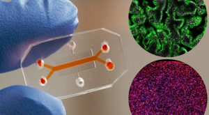 Human intestinal chip microfluidic culture device
Reference:
[1] Kim H J, et al. Human gut-on-a-chip inhabited by microbial flora that experiences intestinal peristalsis-like motions and flow[J]. Lab on a Chip, 2012, 12(12): 2165-2174.
[2] Xiang Y Q, et al. Construction of gut-on-a-chip based on bio-microfluidic technology[J]. Journal of Integration Technology, 2020, 9(3): 56-65.
[3] Kim H J, et al. Gut-on-a-Chip microenvironment induces human intestinal cells to undergo villus differentiation[J]. Integrative Biology, 2012,5(9):1130-1140.
Related Services: