With the continuous innovation and development of organ-on-a-chip technology, its applications in physiological research, drug screening, disease model establishment, environmental assessment, etc. are becoming more and more extensive, and the products are becoming more and more abundant. The development of multiple organ-on-a-chip provides an innovative means of drug development and evaluation for the study of the complexity, functional changes, and integrity of the body’s organs. Organ-on-a-chip technology not only simplifies the general drug development process, but also provides a more clinically homogeneous model for drug safety evaluation.
As an enterprise specialized in the research and development of organ chips, Creative Biolabs provides comprehensive and high-quality organ chip-based detection services, which can observe the growth status of different cells at different times and spaces, and detect various corresponding indicators.
Detection Technology Based on Organ Chips
Mitochondrial potential assay
Mitochondria in cells play a key role in studying disease progression and drug toxicity in organ-on-a-chip models. Therefore, real-time monitoring of mitochondrial potential can provide critical information on the development of mitochondrial dysfunction and the loss and gain of mitochondrial function. Changes in mitochondrial membrane potential are one of the main indicators for evaluating mitochondrial function. In recent years, fluorescent dyes (generally lipophilic cationic compounds) that measure mitochondrial membrane potential have been recognized as common tools for monitoring changes in important mitochondrial parameters. In the presence of transmembrane potential, these dyes bind to the mitochondrial matrix, and the increase or decrease in fluorescence observed by fluorescence microscopy can reflect the increase or decrease in the electronegativity of the mitochondrial inner membrane.
- TMRM method
TMRM is a cell-permeable cationic fluorescent dye with a single laser excitation and a single fluorescence emission peak. Dye diffusion occurs in damaged mitochondria with reduced membrane potential. The advantages of this method are the low rate of organelle binding, no aggregates formed by the dyes, and weak interaction with membrane proteins. The dyeing time is about 30-60 minutes.
- JC-1 method
JC-1 is a widely used small molecule fluorescent probe of mitochondrial membrane potential, which accumulates in mitochondria along with mitochondrial membrane potential. When the membrane potential is high, JC-1 aggregates in the matrix and produces red fluorescence (590nm); when the membrane potential is low, JC-1 cannot aggregate but exists as a monomer, and the ratio of red/green fluorescence intensity decreases, resulting in green fluorescence (530nm). Therefore, changes in mitochondrial membrane potential can be detected quickly and easily by fluorescence color changes. The dyeing time is about 10-60 minutes.
Drug Toxicity Study
During the drug development process, drugs entering clinical trials must undergo toxicity studies to ensure that they are safe before being administered to humans. The in vitro model constructed using organ chip technology can simulate the microtissue of human organs, and can detect and characterize the specific toxic reactions induced by various candidate drugs or compounds in different organs with high throughput, which has an important application prospect in drug development and toxicity research.
- Hepatotoxicity Evaluation
The liver plays the roles of deoxidation, storage of glycogen, and secretory protein synthesis in the body. It is also the main site for the biotransformation and detoxification of substances, and is an important target organ for drug toxicity. Drug-induced liver injury is an important cause of acute and chronic liver diseases, and it is also the main reason for the failure of new drug development. Therefore, performing hepatotoxicity testing is a critical step in drug development. Multi-cell cultured liver chips with specific species (such as humans, dogs, and rats) can be constructed for the hepatotoxicity test of drugs, and the detection range includes liver cell damage, cholestasis, Kupffer cell exhaustion, and steatosis. In addition, it is also possible to simulate and determine the correlation between humans and animals in drug hepatotoxicity testing.
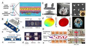 Liver-on-a-chip system for drug toxicity testing
- Renal Toxicity Evaluation
Drug nephrotoxicity is a more serious type of drug toxicity, accounting for about 25% of serious adverse drug reaction cases in clinical settings. In recent years, the kidney-on-a-chip model has gradually been used to test drug nephrotoxicity due to its high bionicity, in which the presence of fluid shear stress makes cells more sensitive to drugs, and the use of primary cells makes in vitro nephrotoxicity tests and in vivo results more physiologically relevant. The kidney-on-a-chip model can realize co-culture of various cells, in vivo microenvironment simulation, and in vitro biomarker analysis.
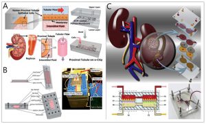 Kidney-on-a-chip system for drug toxicity testing
- Cardiotoxicity Evaluation
Cardiotoxicity refers to the direct damage caused by the drug to the heart tissue and the indirect damage caused by the effect of the drug on the hemodynamics, such as causing cardiac thrombosis. Preclinical screening of drug cardiotoxicity is an important step in the development of new drugs. Traditional models cannot accurately predict whether drugs are cardiotoxic to humans. The development of microfluidic technology has made the heart chip a new type of in vitro tool. The platform can simulate the blood circulation system and connect various tissue cells to realize the co-cultivation of human healthy heart cells and pathological cells in different chambers. Adverse drug reactions can also be simulated using integrated pneumatic valves and pumps to generate precise fluid flows.
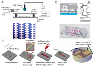
Heart-on-a-chip system for drug toxicity testing
Neural Activity Analysis
The organ-on-a-chip platform represented by Creative Biolabs can provide an accurate and controllable microenvironment for neuronal cell culture. Neuron cell protrusions have plasticity, and neurons are seeded in microchannels through the network array microstructure, so that cell protrusions grow along the microchannels due to physical space structure constraints, and the protrusions are connected to each other to form a neuron network.
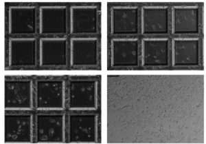 Culture of neuronal networks in chips at different cell densities
Calcium ions are important intracellular signaling molecules in the mammalian nervous system. When neurons are in an excited state, the concentration of intracellular calcium ions can increase by 10 to 100 times, that is, the activity of neurons is closely related to the concentration of calcium ions inside, and neurons will burst into a short-term calcium ion concentration when they discharge. peak. Therefore, with the help of the corresponding relationship between calcium ion concentration and neuron activity, special fluorescent dyes such as calcium indicator (Fluo-4) are used to express the calcium ion concentration in neurons through fluorescence intensity, so as to analyze the neuron activity, i.e., the neuronal calcium imaging technology.
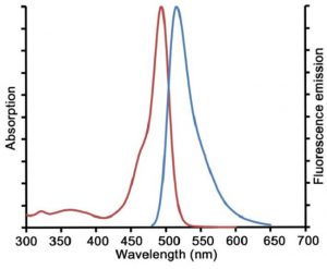 Excitation and emission spectra of Fluo-4
Reference:
[1] Jang K J, et al. Reproducing human and crossspecies drug toxicities using a Liver-Chip[J]. Sci Transl Med, 2019, 11(517): 5516.
[2] Cong Y, et al. Drug Toxicity Evaluation Based on Organ-on-a-Chip Technology: A Review [J]. Micromachines (Basel), 2020, 15(23): 20-28.
[3] Brana I, Tabernero J. Cardiotoxicity[J]. Ann Oncol, 2010, 21(7): vii173-9.
[4] Kamei K I, et al. Integrated heart /cancer on a chip to reproduce the side effects of anti-cancer drugs in vitro[J]. RSC Adv, 2017,7(58): 36777-36786.
Related Services: