Blood-Brain Barrier
The blood-brain barrier (BBB) is a highly selectively semipermeable border of endothelial cells that prevents the nonselective passage of solutes from circulating blood into the extracellular fluid of the central nervous system, where neurons reside. The BBB is formed by the endothelial cells of the capillary wall, the ends of the astrocytes wrapping the capillaries, and the pericytes embedded in the capillary basement membrane. The BBB can control the transport of substances in the blood and brain tissue and also plays a key role in environmental homeostasis and drug transport in the brain.
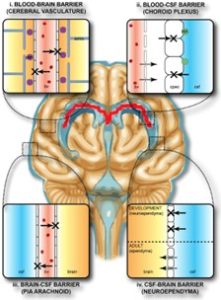 Figure 1. The protective barrier of the brain (collectively known as the blood-brain barrier).
Blood-brain barrier chip model
At present, most studies rely on animal models or in vitro cell culture models, which are difficult to dynamically control the microenvironment of cell culture, including the effects of fluid flow, shear stress strain, and mechanical compression at the physiological level. The establishment of the BBB chip model based on microfluidics overcomes these problems and can simulate the in vivo microenvironment of the BBB to the greatest extent. The BBB chip refers to an in vitro BBB model system that is easy to regulate and close to the in vivo microenvironment constructed by using microfluidic technology and three-dimensional cell culture technology. The device, which consists of a vascular lumen and a ventricle separated by a porous membrane, can achieve similar in vivo microenvironmental and structural functions by manipulating the geometric, mechanical, and biochemical properties of the environment. It can be used to establish models related to brain diseases and evaluate the efficacy and toxicity of drugs, etc.
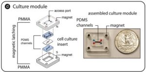 Figure 2. Blood-brain barrier-organ-on-a-chip model.
Features and advantages of the blood-brain barrier chip model
- Low manufacturing cost, flexible design
- Smaller functional volume, less cell number required
- Ability to provide a parallel and controllable dynamic brain microenvironment
- Fast media exchange to reach a steady state quickly
- Allowing permeability determination and controlled delivery of test substances
- Allowing inspection of cell viability by microscopy
- Ability to be integrated into biosensors for long-term monitoring of blood-brain barrier function
Detection based on blood-brain barrier-organ chip model
Based on the BBB chip model, a variety of BBB functional studies can be completed in vitro, predicting the permeability of drugs through the human BBB in healthy or diseased states, evaluating the efficacy of drugs passing through the BBB into brain tissue and BBB-related neurodegenerative diseases or basic research on diseases such as brain tumors. Creative Biolabs represents a variety of detection services based on the BBB organ chip model. These detection technologies include but are not limited to:
1. High-content imaging analysis
High-content imaging analysis refers to understanding cell changes and cell activities through automated cell imaging analysis using cells as the detection object under the premise of maintaining the integrity of cell structure and function. High-content imaging technology can perform fully automatic high-throughput imaging analysis on in vitro BBB chip models, and provide high-definition images, including single cell images, statistical analysis results of cell populations, changes in cell number and morphology, and subcellular structure, as well as analysis of multiple indicators such as changes of the fluorescent signal over time and spatial distribution, etc.
High-content analysis is generally divided into three processes:
- Monitor cells using single or multiple fluorescent probes.
- Quickly capture pictures of cells with imaging equipment for high-resolution images.
- Extract detailed information from the pictures for data analysis using various software.
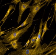 Figure 3. Schematic diagram of high-content analysis.
2. Blood-brain barrier integrity and permeability analysis
The molecular basis of BBB function depends on tight interactions between adjacent brain microvascular endothelial cells (BMECs). Measurement of transmembrane electrical resistance (TEER) and permeability analysis are commonly used methods to evaluate BBB integrity. It integrates electrode materials into the BBB chip for real-time measurement, and tracks and evaluates the integrity and tightness of the cell layer in the in vitro BBB model. Permeability assays quantify barrier permeability by quantifying molecules that penetrate the BBB using fluorescently labeled molecular tracers. By using fluorescent tracers of different molecular weights, the quality of tight junctions can be assessed.
Specific detection technology:
- Permeability markers. Use appropriate control compounds to assess cell monolayer integrity and to validate experimentally determined transport properties of drugs or other compounds.
- Blink counting. Add a radiolabeled substance to a diffusion chamber, determine its permeability, and then evaluate the effect of the unlabeled compound on the permeation of the radiolabeled compound.
- Capillary Electrophoresis (CE). CE can solve some research projects that have specific requirements in terms of sample volume.
- Liquid Chromatography/Mass Spectrometry. Peptide quantification studies using liquid chromatography/mass spectrometry can determine the transport of specific compounds on BMEC monolayers.
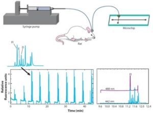
Figure 4. Detection of BBB integrity and excitability by microdialysis sampling-coupled chip electrophoresis.
3. Blood-brain barrier transport research
Drugs and other substances pass through the BBB mainly through four pathways, namely:
- Small molecules diffuse through the intercellular space;
- Fat-soluble molecules melt and diffuse;
- Specific receptor-mediated swallowing;
- Activation of specific carrier channels and enzyme systems.
The research on the transport mechanism of BBB not only helps to understand the pathophysiological changes of brain capillary endothelial cells and surrounding parenchymal cells, but also has very important guiding significance in drug discovery, delivery, and targeting. Transport studies based on the BBB organ-on-a-chip model include:
- Screening for efflux carrier inhibitors. The existence of the efflux mechanism on the BBB has led to the efflux of many chemotherapeutic drugs. Finding drugs that can effectively inhibit the efflux carrier is helpful for the uptake of drugs and the reversal of related drug resistance.
- Carrier-mediated drug transport studies. Nutrients in the blood circulation can cross the BBB and enter the brain tissue by means of a carrier-mediated transport system. By substrateizing the drug, it enters the brain tissue together with nutrients.
- Receptor-mediated drug transport studies. Some endogenous macromolecules such as insulin can be recognized by the corresponding receptors on the luminal surface of the capillary endothelium in the brain, and enter the brain tissue through transcytosis. Ligands can be designed and optimized by bioengineering technology and used as carriers for drug delivery.
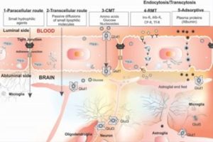
Figure 5. Different mechanisms by which drugs enter the brain from the blood.
4. Neurotoxicity detection based on BBB chip
Some pathological factors can induce the activation of matrix metalloproteinases, resulting in decreased cell viability, decreased tight junctions between vascular endothelial cells, basement membrane degradation, and BBB integrity damage. As a result, macromolecules and harmful substances can enter the brain tissue and cause different degrees of brain damage. Services such as cell viability assays, cytotoxicity assays, and permeability assays allow the investigation of the effects of drugs and other factors on the integrity of the BBB. The detection range includes:
- Assessment of barrier structural damage due to neurotoxicity. Many neurotoxins can directly disrupt the structure of the barrier and increase its permeability, allowing the poison to come into contact with the brain parenchyma. It is characterized by symmetrical vacuolation and microvacuolation in selected brain regions.
- Detection of neurotoxicity due to altered barrier function. Some chemical substances, such as aluminum and lead, will not directly destroy the shape of the barrier, but will change its regulatory function, leading to brain homeostasis disorders and neurotoxicity.
- Biotransformation studies of the brain barrier. Brain endothelial cells and choroidal epithelial cells contain a variety of drug-metabolizing enzymes, and differences in BBB metabolism in different species can alter the way that some specific compounds are present. For example, biotransformation of 1-methyl-4-phenyl-1,2,3,6-tetrahydropyridine (MPTP) causes Parkinson’s syndrome in humans and primates.
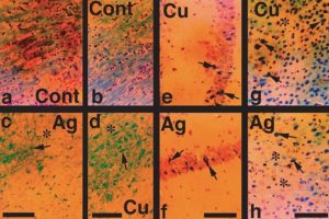 Figure 6. Rat nerve cell injury detection.
5. Research on the interaction between BBB and the immune system
Some studies suggest that aberrant immune activation may be due to BBB disruption, which exposes normally immunosegregated central nervous system (CNS) antigens to the systemic immune system, inducing autoimmune responses that lead to disease. In addition, the immune response also plays an important role in neurological diseases such as neurodegenerative/degenerative diseases. Research areas include:
- To study the interaction between BBB and T cells
In the BBB organ chip, endothelial cells and astrocytes participate in the restriction, antigen recognition, and presentation of immunocompetent cells in the brain by expressing adhesion molecules and other cell surface antigens, and play a complex and important role in CNS immune regulation. For example, activated T cells cross the BBB into the brain parenchyma and play an important role in the pathogenesis of CNS immune diseases such as multiple sclerosis (MS).
- Study the role of BBB in participating in antigen presentation
The activated lymphocytes transferred from the peripheral vein to the CNS can become specific effector cells, which indirectly shows that the BBB participates in the antigen presentation of the CNS. The BBB controls the number and type of lymphocytes entering the CNS through the antigen recognition process between adhesion molecules expressed by endothelial cells and lymphocytes. BBB microvessels and capillary endothelial cells are not only closely connected with microvascular pericytes, smooth muscle cells, and astrocytes, but also some cells communicate with each other through gap junctions. Additionally, some adhesion molecules are expressed in endothelial cells and astrocytes, thus providing a structural basis for the presentation of antigens and the transmission of immune signals.
- Studying peripheral antibodies through the BBB and brain immune injury
Neuroimmunology has done a lot of research on the role of the BBB in brain immune injury, especially in the pathogenesis of MS. Activated T lymphocytes can cross the BBB through cell surface proteins such as adhesion molecules expressed by the endothelial cells of the BBB and enter the brain to mediate a series of immune responses. Related studies have also proved that peripheral high levels of antibodies against common antigens of brain components also play an important role in the occurrence of brain immune diseases. When the BBB permeability increases to a certain extent, peripheral antibodies can cross the BBB and enter the brain tissue, resulting in brain immune damage.
References:
[1] Daneman R, Prat A. The blood-brain barrier. Cold Spring Harb. Perspect. Biol. 2015, 7(1): a020412.
[2] Ballabh P, et al. The blood-brain barrier: an overview: structure, regulation, and clinical implications. Neurobiol Dis. 2004, 16(1): 1–13.
[3] Helen B S, et al. Immune responses at brain barriers and implications for brain development and neurological function in later life. Front. Integr. Neurosci. 2013, 7: 61.
[4] Kaisar, M A, et al. In Vitro BBB Models: Working with Static Platforms and Microfluidic Systems. Blood-Brain Barrier. 2019, 142.
[5] Kuhnline S C, et al. Analytical and biological methods for probing the blood-brain barrier. Annul. Rev. Anal. Chem. 2012, 5: 505-531.
[6] Barar J, et al. Blood-brain barrier transport machineries and targeted therapy of brain diseases. BioImpacts. 2016, 6(4): 225.
[7] Sharma H S, et al. Influence of engineered nanoparticles from metals on the blood-brain barrier permeability, cerebral blood flow, brain edema and neurotoxicity. An experimental study in the rat and mice using biochemical and morphological approaches. J. Nanosci. Nanotechnol. 2009, 9(8): 5055-5072.
Related Services: